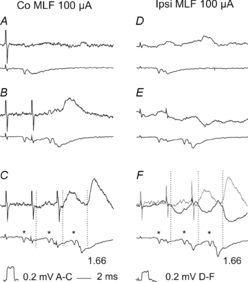
Figure 3. Examples of disynaptic EPSPs evoked from the contralateral MLF and disynaptic IPSPs from the ipsilateral MLF in CC DSCT neurones.

Upper traces in A–C and in D–F are from two CC DSCT neurones located in the left and right Clarke's column, respectively, recorded in the same experiment after the left hemisection of the spinal cord. Lower traces are from the surface of the spinal cord. Averages of 40 records. Dotted lines indicate onset of PSPs evoked by stimuli applied in the right MLF, with latencies given from the first components of the descending volleys (*). Grey trace in F, records from C. Note that EPSPs and IPSPs were evoked at similar latencies (1.66 ms after the 3rd stimulus) compatible with disynaptic coupling via single excitatory or inhibitory interneurones, and that peak amplitudes of PSPs evoked after subsequent stimuli increased.
