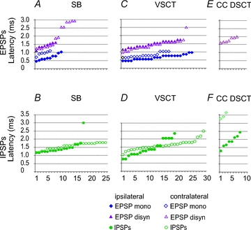
Figure 4. Comparison of latencies of earliest components of EPSP and IPSP evoked from the MLF in three populations of spinocerebellar neurones.

A and B, C and D and E and F, latencies of PSPs (ordinate) evoked in samples of SB, VSCT and CC DSCT neurones (abscissa). In each plot filled symbols are for PSPs evoked from the ipsilateral MLF after contralateral hemisection of the spinal cord (i.e. via ipsilaterally descending MLF fibres) and open symbols are for those evoked after ipsilateral hemisection (i.e. via contralaterally descending MLF fibres). Data for ipsilateral actions on VSCT neurones are estimated from the histogram of latencies of PSPs in Fig. 4 in Baldissera & Roberts (1975). All PSPs were evoked by stimuli ≤100 μA. When both monosynaptic and di- or trisynaptic PSPs were evoked, latencies of both were measured separately. The latencies are ranked from the shortest to longest. Note that in CC DSCT neurones no EPSPs were evoked from the ipsilateral MLF and that only di- or trisynaptic EPSPs followed stimuli applied in the contralateral MLF in contrast to SB and VSCT neurones in which both mono- and disynaptic EPSPs were evoked from the ipsilateral as well as the contralateral MLF. Note also that a considerable proportion of EPSPs and IPSPs were evoked at similar latencies via ipsilaterally or contralaterally descending MLF fibres.
