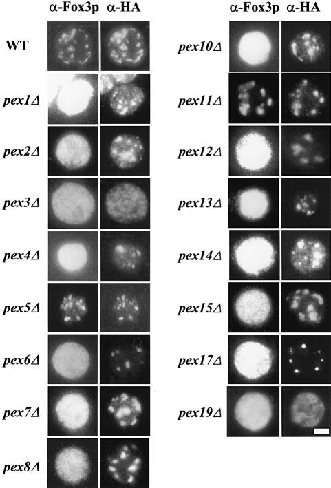
Fig. 1. pex3Δ and pex19Δ cells lack Pex11p–HA-containing membrane structures. Oleic acid-induced wild-type and pexΔ mutant cells expressing HA-tagged Pex11p as a marker for peroxisomal membranes were processed for double immunofluorescence microscopy using rabbit polyclonal antibodies specific for peroxisomal thiolase (Fox3p) and mouse monoclonal antibodies specific for the HA epitope. Secondary antibodies were CY3-conjugated anti-mouse IgG and FITC-conjugated anti-rabbit IgG. Bar, 5 μm.

An official website of the United States government
Here's how you know
Official websites use .gov
A
.gov website belongs to an official
government organization in the United States.
Secure .gov websites use HTTPS
A lock (
) or https:// means you've safely
connected to the .gov website. Share sensitive
information only on official, secure websites.
