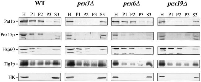
Fig. 3. pex3Δ and pex19Δ cells mislocalize their PMPs to the cytosol. Subcellular distribution of PMPs and marker enzymes in oleic acid-induced wild-type and pexΔ mutant cells. After subcellular fractionation equivalent volumes of the 600 g post-nuclear supernatant [homogenate (H)], 2500 g pellet (P1), 25 000 g pellet (P2), 150 000 g pellet (P3) and 150 000 g supernatant (S3) were analysed by immunoblotting. For detection of the PMPs Pat1p and Pex15p in pex3Δ and pex19Δ cells, samples were concentrated 10–fold by trichloroacetic acid precipitation before loading. Antibodies were directed against the proteins as indicated. Cytosolic marker, hexokinase (HK). Mitochondrial and endosomal membrane markers are Hsp60 and Tlg1p, respectively. PMPs are Pat1p and Pex15p.

An official website of the United States government
Here's how you know
Official websites use .gov
A
.gov website belongs to an official
government organization in the United States.
Secure .gov websites use HTTPS
A lock (
) or https:// means you've safely
connected to the .gov website. Share sensitive
information only on official, secure websites.
