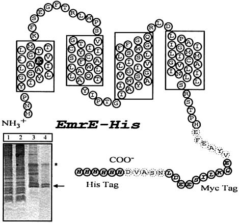
Fig. 1. Model of EmrE–His with the four transmembrane regions predicted by hydropathy plots. The His and Myc tags are indicated by open ovals with one-letter amino acid codes shown in bold italicized text. Dashed ovals indicate residues that were incorporated into the transporter in the process of linking in the epitope tags. The Glu14 residue in the first transmembrane region is highlighted. The Myc tag is used here only as a linker to keep the His residues away from the membrane. Without the linker, the His-tagged transporter displays only residual activity. Inset, SDS–PAGE analysis of different stages of purification. Lane 1, total membranes; lane 2, detergent-solubilized extract after Ni–NTA purification, unbound fraction; lane 3, EmrE after Ni–NTA purification; lane 4, EmrE after size exclusion purification. The arrow indicates the monomeric form of EmrE; the asterisk the dimeric form. The apparent Mr of monomeric EmrE–His is 14 400 Da.

An official website of the United States government
Here's how you know
Official websites use .gov
A
.gov website belongs to an official
government organization in the United States.
Secure .gov websites use HTTPS
A lock (
) or https:// means you've safely
connected to the .gov website. Share sensitive
information only on official, secure websites.
