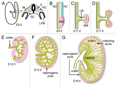
Figure 1.
Schematic diagram of development of the renal collecting duct system. (A) Diagram illustrating location of intermediate mesoderm (IM), including the nephric duct (ND) and nephrogenic cord (NC) in the mouse embryo. S, somite; LPM, lateral plate mesoderm. (B) Formation of metanephric mesenchyme (MM). (C) Outgrowth of ureteric bud (UB). (D) First branching of the UB. (E and F) Continued UB branching and formation of the nephrogenic zone (dark pink). (G) Elongation of collecting ducts to form the medulla. Modified from reference 13.

