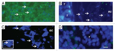
Figure 5.
Fluorescence microscopy of tissue sections originating from (A) a normal porcine pancreas or (B–D) a diabetic rat that had been transplanted with embryonic pig pancreas in mesentery and subsequently with porcine islets in kidney: (B) a subcapsular section from kidney, T tubule, RC Renal Capsule; (C) a section of mesenteric lymph node, GC, germinal center, INSET enlargment; and (D) renal cortex, T, tubule. Arrows (A–C) delineate pig X chromosomes. Scale bar 10 um (D). Reproduced with permission from the American Society for Investigative Pathology.12

