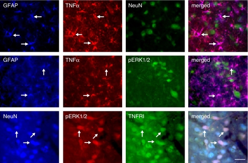
Figure 8.
Determination of cellular localization of TNFα, TNFRI, pERK1/2, Fos, and pCREB in the vlPAG. These rats were injected with control vector T0Z before the morphine withdrawal protocol. All labeled proteins are endogenous proteins. Triple-label immunostaining of GFAP, TNFα, and NeuN in the vlPAG (upper panel). There was an almost complete colocalization between GFAP (blue) and TNFα (red) imaging, but TNFα did not colocalize with NeuN (green), which suggested that TNFα located on astrocytes, but not neurons. Immunostained pERK1/2 did not colocalize with either GFAP or TNFα (middle panel). Triple-label immunostaining (lower panel) showed that NeuN (blue) was colocalized with endogenous TNFRI (green). pERK1/2-positive cells (red) colocalized extensively with both NeuN and TNFRI immunostaining, suggesting that pERK1/2-positive cells be located on TNFRI-positive neurons. Arrows indicate double/triple-labeled cells. Scale bar, 100 μm.

