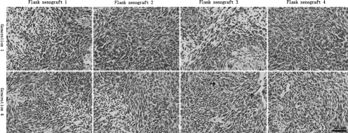
Figure 3.
Histopathologic features of flank xenografts. Compared with the human surgical materials, GBM xenografts in 4 different xenograft lines displayed pleomorphic cells, presence of mitotic activity, necrotic foci, and mild microvasculars (black arrow), but endothelial proliferation with multilayering of endothelial cells was not observed.

