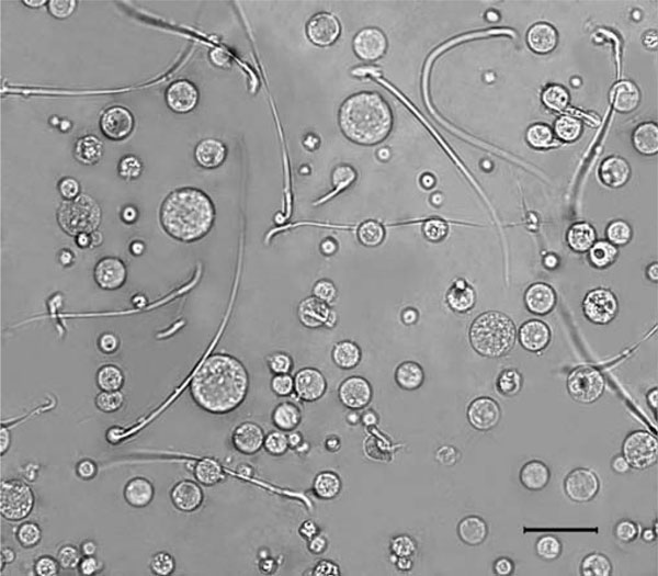
Figure 1.
Partial view of a cell suspension from adult rat testis. The suspension was prepared with the Medimachine as described in the "Materials and methods" section and visualized by phase contrast microscopy. The wide variety of cell sizes and shapes can be observed in a well-dissagregated state. As can be seen, most of the spermatozoa keep their flagellae. The absence of multinucleates and the integrity of cell cytoplasms are also evident. The bar corresponds to 25 μm.

