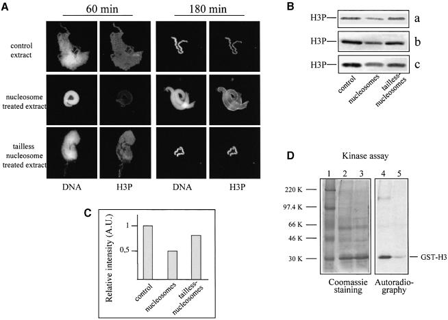
Fig. 7. A kinase activity phosphorylating histone H3 at Ser10 was found associated with nucleosomes. (A) Nucleosome-arrested chromosome condensation exhibits a specific pattern of histone H3 phosphorylation. Sperm nuclei were incubated for 60 or 180 min in mitotic extract, containing either native or tailless nucleosomes, and DNA and histone H3 phosphorylation were visualized by Hoechst 33258 and anti-phosphorylated histone H3 antibody. (B) Native nucleosomes present in the extract decrease the degree of histone H3 phosphorylation of the chromosome condensation intermediates. Extracts were incubated for 30 min with native or tailless nucleosomes, the nucleosomes were pelleted by centrifugation and the supernatants supplemented with sperm nuclei. The control and the two nucleosome-treated extracts were incubated for 180 min at room temperature and the remodelled sperm were pelleted by centrifugation. The proteins from the pellets were run on an SDS–18% polyacrylamide gel, and phosphorylated histone H3 was detected with a specific antibody; a, b and c represent the results of three independent experiments. (C) Quantification of the data presented in (B). (D) The pelleted native nucleosomes are associated with kinase activity phosphorylating histone H3. The pellets from the control and nucleosome-treated extracts (see above) were resuspended and supplemented with GST–H3 and [γ–32P]ATP. After 30 min incubation, the samples were precipitated with TCA and analysed on an SDS–12% polyacrylamide gel. Left, Coomassie staining of the gel; right, autoradiography. Lane 1, protein marker; lane 2, control pellet; lane 3, pellet from the nucleosome-treated extract; lanes 4 and 5, autoradiography of lanes 3 and 4, respectively. The positions of GST–H3 and the protein markers are indicated.

An official website of the United States government
Here's how you know
Official websites use .gov
A
.gov website belongs to an official
government organization in the United States.
Secure .gov websites use HTTPS
A lock (
) or https:// means you've safely
connected to the .gov website. Share sensitive
information only on official, secure websites.
