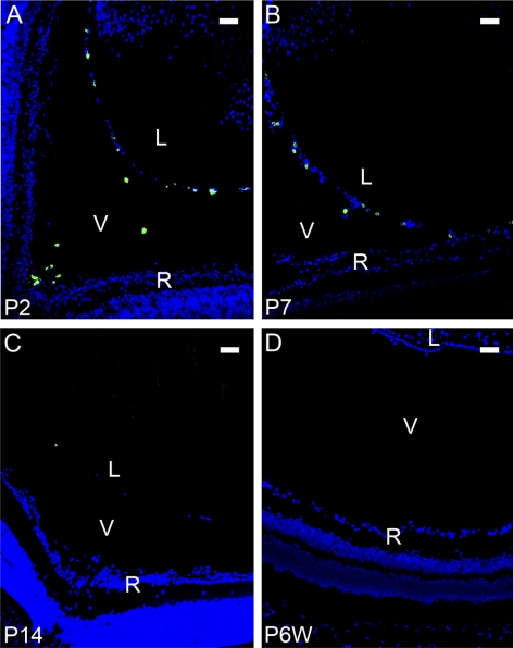
Figure 2.
LYVE-1 expression with the regression of the HVS at postnatal stages. Representative cross-sectional micrographs demonstrating LYVE-1 expression at postnatal stages, which was obvious at P2 (A) and P7 (B), barely detectable at P14 (C), and completely absent in the adult (D). Green: LYVE-1; blue: DAPI nuclei staining. L, lens; V, vitreous; R, retina. Scale bars, 50 μm.

