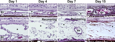
Figure 2.
Light micrographs of transverse sections through the sclera-choroid-RPE of control and treated chicken eyes after 1, 4, 7, and 15 days of recovery from form-deprivation myopia. Note the expansion of the choroid and the appearance of thin-walled lymphatic channels on the scleral side of the choroid (asterisks). Erythrocytes can be seen in large and small blood vessels (BV). Scale bar, 50 μm.

