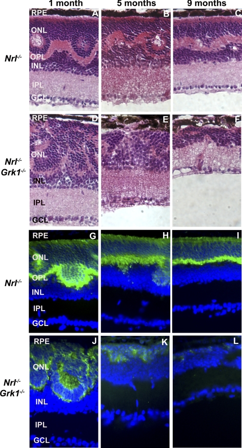
Figure 2.
Age-dependent cone photoreceptor degeneration in the Nrl−/− and Nrl−/−Grk1−/− mouse retina. Retinal sections were (A–F) stained with H&E or (G–L) immunostained with anti–mCAR-LUMIJ, followed by a fluorescein-conjugated anti–rabbit IgG secondary antibody. Significant thinning of the ONL is observed at 5 months, with complete loss of cones by 9 months. Photographs were taken from the central inferior region of the retina.

