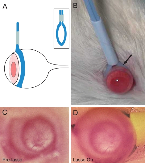
Figure 2.
Schematic (A) and photograph (B) of the adjustable vascular loop placed around an eye of a Sprague-Dawley rat posterior to the limbus. Retinal vasculature at the optic disc appears unchanged before (C) and while (D) the vascular loop is placed around an eye completing 6 weeks of daily elevated IOP.

