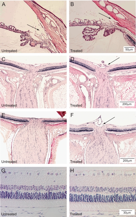
Figure 4.
Histologic sections of the anterior chamber angles of one 6-week-treated rat with the untreated eye (A) and treated eye (B) for comparison (hematoxylin and eosin; scale bar, 50 μm). No evidence of synechiae is present in the angle. Histologic sections of the retina and optic nerves of one 6-week-treated rat with the untreated eye (C) and treated eye (D) for comparison. Retinal ganglion cells are present in both specimens. No evidence of ischemic injury is present (hematoxylin and eosin; scale bar, 200 μm). A suggestion of increased optic nerve cupping is present in the treated eye (black arrow). Histologic sections of the retina of another rat with the untreated eye (E) and treated eye (F) for comparison demonstrates similar findings (hematoxylin and eosin; scale bar, 200 μm). A suggestion of increased optic nerve cupping is present in the treated eye (black arrow). Retinal layers are intact in both untreated (G) and treated (H) eyes. No evidence of ischemic injury is present (hematoxylin and eosin; scale bar, 30 μm).

