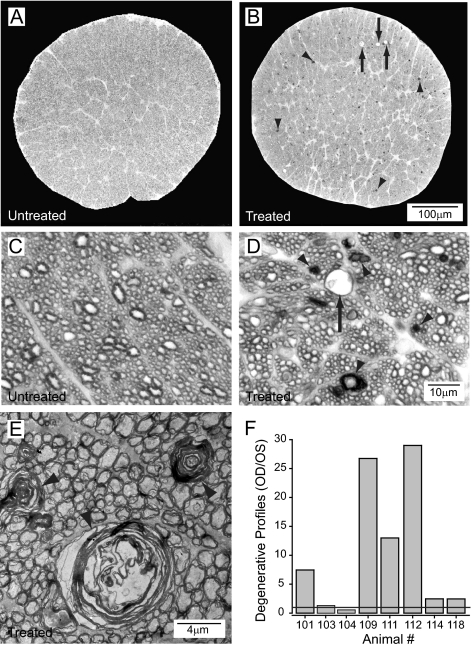
Figure 6.
Representative semithin rat optic nerve cross-sections (toluidine blue) of an untreated eye (A, C) and the contralateral 6-week treated eye (B, D). More alterations of axonal structures are present in the treated side (B, D) with degenerated axons (arrowheads) and vacuoles (arrows) compared with structures in the untreated side (A, C). (E) Myelin wrapping abnormalities (arrowheads) in the optic nerve are evident in an electron micrograph of a 6-week treated eye. (F) More degenerating profiles are observed in optic nerve cross-sections in treated compared with untreated contralateral eyes. Although treatment was uniform, some animals had a greater degenerative response than other individuals.

