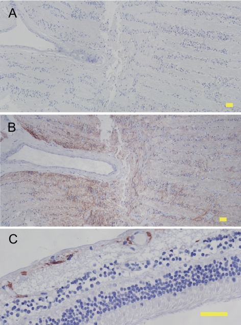
Figure 5.
IL-8 protein expression in human cadaver eyes. Immunohistochemistry was performed on sections of paraformaldehyde-fixed human cadaver eyes using negative control IgG (A) or anti–IL-8 antibody (B, C) and detection with Nova Red substrate followed by hematoxylin counterstain. Reddish-brown: positive immunoreactivity. Blue: hematoxylin counterstain. Staining with negative control IgG resulted in no detectable immunoreactivity (A). IL-8 expression was found in the optic nerve (B) and inner retina (C). Yellow scale bar, 50 μm.

