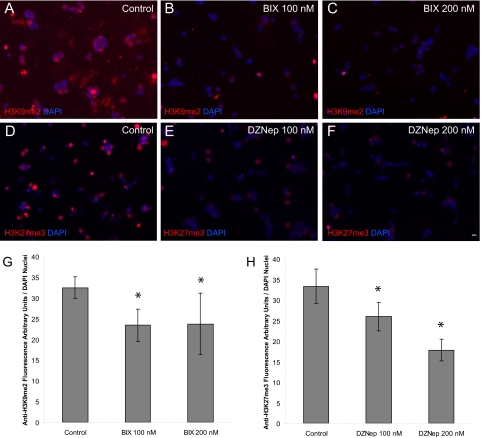
Figure 4.
BIX-01294 (BIX) and DZNep inhibit H3K9me2 and H3K27me3 in P0 RGCs. (A–C) Immunofluorescence detection of H3K9me2 (red) and (D–F) H3K27me3 (red) with or without treatment of the described concentrations of BIX and DZNep was overlaid on the nuclear counterstain (DAPI, blue). Scale bar, 5 μm. (G) P0 RGCs were cultured for 3 days and treated with vehicle control, 100 nM and 200 nM BIX, or the same concentrations of (H) DZNep. Relative fluorescence per nuclei was obtained by dividing the fluorescence signal (arbitrary units) of the corresponding histone mark antibody by the total number of DAPI nuclei per 20× field using Image J software (mean ± SEM; n = 2 for each treatment group, eight fields per culture, mean 612 nuclei counted per field. *P < 0.008.

