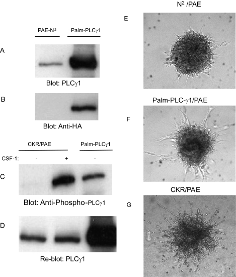
Figure 1.
Selective activation of PLCγ1 induces tube formation by endothelial cells. An equal number of serum-starved PAE cells expressing an empty vector (PAE-N2) or constitutive active HA-tagged PLCγ1 (palm-PLCγ1) were lysed, and Western blot analysis was performed to determine total PLCγ1 using anti-PLCγ1 (A) or anti-HA (B) antibody. PAE cells expressing chimeric VEGFR-2 (CKR) were unstimulated or stimulated with CSF-1 for 10 minutes, and the cells were lysed. PAE cells expressing palm-PLCγ1 were lysed without stimulation. Cell lysates were subjected to Western blot analysis with a phospho-specific PLCγ1 antibody (C). The same membrane was analyzed for total PLCγ1 levels (D). PAE cells expressing the empty vector PAE-N2, palm-PLCγ1, or chimeric VEGFR-2 (CKR) were subjected to tube formation assay, and photographs were taken after 48 hours (E, F, G).

