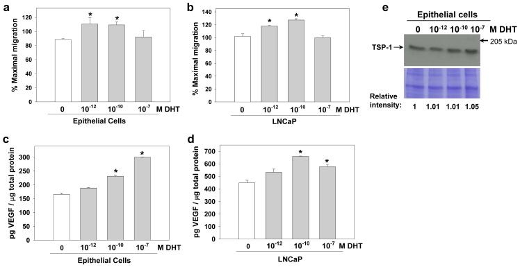
Figure 1.
Androgens induce angiogenic activity in prostate epithelial cells. Serum-free conditioned media were collected from normal prostate epithelial cells (PrEC) and from androgen sensitive LNCaP PCa cells treated with DHT (0-10−7 M). (a,b) Angiogenic activity was assessed in PrEC and LNCaP conditioned media (20 μg/ml) using a microvascular endothelial cell migration assay, and data are presented as the mean ± SE of the percent maximum migration toward the positive control. (c,d) VEGF levels were measured in conditioned media by ELISA. Data are presented as mean ± SD of two replicates. (e) TSP-1 expression was evaluated by Western blot. The lower panel is an identically loaded gel to show equal protein loading between lanes. Protein levels relative to untreated (0 DHT) were determined by densitometry. Protein size standard is indicated. *Significantly different compared to the untreated cells within each experiment, P<0.05.

