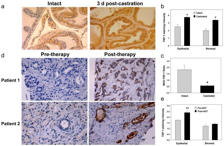
Figure 5.
TSP-1 expression increases following androgen ablation in the mouse prostate and in patient-matched biopsy specimens in vivo. Prostate tissue sections were immunostained with anti-TSP antibody. (a) Prostate tissues were collected from intact or castrated (1-21 days post-castration; n=5 per group) wildtype (C57Bl/6) mice. Sections from intact and 3 days post-castration are shown. (b) TSP-1 staining intensity was graded on a 1-4 scale in the epithelium and stroma of the dorsal prostate. (c) The vessels were counted in the dorsal prostate stroma and the MVD:TSP-1 staining ratio calculated. (d) Patient-matched biopsies were taken from PCa patients before and 3-7 days after androgen ablation therapy. Six of eight patient tissues showed increased immunoreactivity following androgen ablation therapy. Pre- and post-therapy tissues are shown for two patients. (e) TSP-1 staining was graded on a 1-4 scale in the epithelial and stromal compartments. *Significantly increased in castrated compared to intact, P<0.012, and **significantly increased in human post-ADT tissues, P<00.013.

