Figure 7.
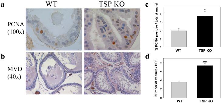
Prostate tissue of TSP-1 KO has increased epithelial cell proliferation and MVD. Mouse prostate tissues were immunostained with anti-PCNA (a) and anti-factor VIII-related antigen (b) to assess proliferation and MVD, respectively. Representative tissue sections are shown. (c) The number of PCNA-positive and the total number of epithelial nuclei were counted at 100X to quantify proliferative activity. A minimum of 150 nuclei were counted for each mouse (n=6 per group). PCNA positivity is presented as a percent of the total number of nuclei counted; *P<0.007. (d) MVD quantified by counting vessels in 5 high power fields (HPF; 40X) for each mouse (n=6 per group; **P<0.00003).

