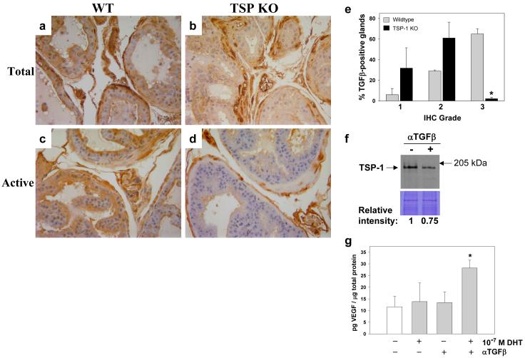
Figure 8.
TSP-1 - TGF-β regulation in the prostate. Wildtype (C57Bl/6) and TSP-1 KO mouse prostate tissues (n=5 per group) were immunostained with antibody against total (a,b) and active TGF-β (c,d). (e) In the dorsal prostate, where staining differences were observed, active TGF-β-positive glands were graded on a 1-3 scale: 1 = light, 2 = medium and 3 = intense staining. The number of grade 3 glands was significantly decreased in the TSP-1 null mice as compared to wildtype (*P<0.0001). (f) Stromal cells were treated with neutralizing TGF-β antibody and TSP-1 levels assessed by Western blot analysis. The lower panel is an identically loaded Coomassie-stained gel to verify equal protein loading. Protein levels relative to the untreated cells was quantified by densitometry. (g) VEGF levels in normal stromal cells treated with DHT ± neutralizing TGF-β antibody VEGF were measured by ELISA. **Significantly decreased compared to untreated cells, P<0.05.

