
Fig. 2.
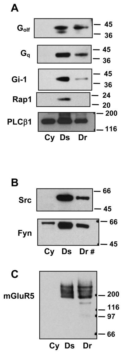
Distribution of signaling molecules into cytoplasmic, detergent soluble and detergent resistant membrane fractions in cortical tissue. A, Components of dopamine-sensitive signaling pathways are primarily localized to Ds and Dr membranes; only PLCβ1 was detected in the cytoplasmic fraction. B, The tyrosine kinases Src and Fyn were associated with Ds and Dr membranes; Fyn, but not Src could be detected in the cytoplasm. C, Another G protein coupled receptor, metabotropic glutamate receptor 5 (mGluR5) distributed into both the Ds and Dr membrane fractions. Twenty micrograms of protein was loaded per lane, except for Dr in panel B, where only 4μg total protein needed to be loaded.