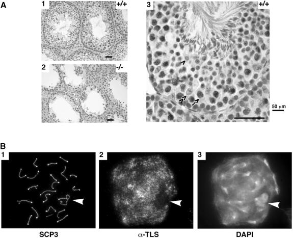
Fig. 3. TLS immunostaining and meiotic progression in wild-type and mutant testes. (A) Formalin-fixed and paraffin-embedded 5 μm sections of wild-type (panels 1 and 3) and mutant testes (panel 2) were reacted with the 4H11 anti-TLS monoclonal antibody, a horseradish peroxidase-conjugated secondary antibody, and the stain developed with diaminobenzidine and counterstained with hematoxylin. Specific cell types in the seminiferous epithelium are identified in panel 3: arrow, pachytene spermatocyte; single arrowhead, round spermatid; double arrowhead, Sertoli cell. (B) Chromosomal spreads of a pachytene/early diplotene spermatocyte fixed and stained for a marker of the axial elements (SCP3), TLS or a chromatin-binding dye (DAPI). The arrowhead points to the sex body that contains the partially synapsed X–Y chromosome pair and is a region of the nucleus with very little TLS staining.

An official website of the United States government
Here's how you know
Official websites use .gov
A
.gov website belongs to an official
government organization in the United States.
Secure .gov websites use HTTPS
A lock (
) or https:// means you've safely
connected to the .gov website. Share sensitive
information only on official, secure websites.
