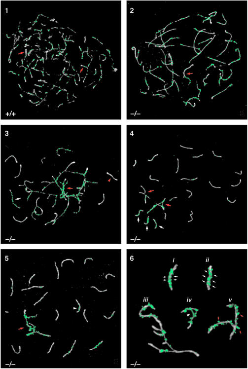
Fig. 4. Analysis of synaptonemal complex formation in TLS–/– spermatocytes by immunolocalization of SCP3 (white) and RAD51 (green) in chromosomal spreads. Panel 1: normal zygotene nucleus from wild-type mouse. Note that synapsis is occurring (red arrows) even as axial elements are still forming. RAD51 localizes to unsynapsed axes and begins to disappear once synapsis has occurred. Panel 2: zygotene nucleus exhibiting synaptic defects. Note the long axial elements that have almost completely formed with little synaptic activity. Some synapsis appears to be non-homologous, based on the differences in length of axial elements (red arrow). Panel 3: zygotene nucleus with major synaptic problems. Synapsed axes show a loss of RAD51 (red arrowhead), while the non-homologously synapsed axes (red arrow) and unsynapsed axes (white arrow) are coated with RAD51. Panel 4: pachytene nucleus with unsynapsed axes and non-homologous synapsis. Considering that the non-homologously synapsed axes involve more than one SC (red arrows), there are >20 axes present in this nucleus. The single axes with a RAD51 coating (white arrows) did not synapse with their homologs. Panel 5: pachytene nucleus containing non-homologously synapsed axes (red arrow). The non-homologously synapsed axes are emphasized by the large amount of RAD51. Also note the abnormal RAD51 bridge connecting two parts of one SC (white arrowhead). Panel 6: high magnification views of abnormal SCs from mutant pachytene nuclei. Incomplete synapsis of two SCs is evident from the double rows of RAD51 along the SCs' lengths (small white arrows) (i and ii). SCP3 staining of these SCs shows that they are thicker than normal, a characteristic of axes that are not fully synapsed. Several axes with partial non-homologous synapses are shown (iii). An abnormal RAD51 bridge has formed between non-homologous regions of the same bivalent (white arrowhead) (iv). SCs containing irregularly shaped RAD51 foci that ‘hang off’ the edges of the SCs, a configuration not observed in normal spermatocytes, are shown (small red arrows) (v).

An official website of the United States government
Here's how you know
Official websites use .gov
A
.gov website belongs to an official
government organization in the United States.
Secure .gov websites use HTTPS
A lock (
) or https:// means you've safely
connected to the .gov website. Share sensitive
information only on official, secure websites.
