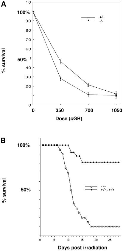
Fig. 5. Increased sensitivity of TLS–/– cells and animals to ionizing irradiation. (A) Day 7 counts of MEFs with the TLS genotypes indicated. The cells were irradiated at day zero with the indicated dose of γ–rays. The number of cells on the plate at day 7 is expressed as a fraction of the cell count on an identical plate at 24 h after plating. Shown are means and SEM of a representative experiment, performed in triplicate and reproduced three times using different pools of MEFs. (B) Survival curve of a cohort of mice discordant for TLS genotype that had received 700 cGR of γ–rays on day 1. Each of the 20 TLS–/– mice was matched with one or more siblings of TLS+/+ or TLS+/– genotype.

An official website of the United States government
Here's how you know
Official websites use .gov
A
.gov website belongs to an official
government organization in the United States.
Secure .gov websites use HTTPS
A lock (
) or https:// means you've safely
connected to the .gov website. Share sensitive
information only on official, secure websites.
