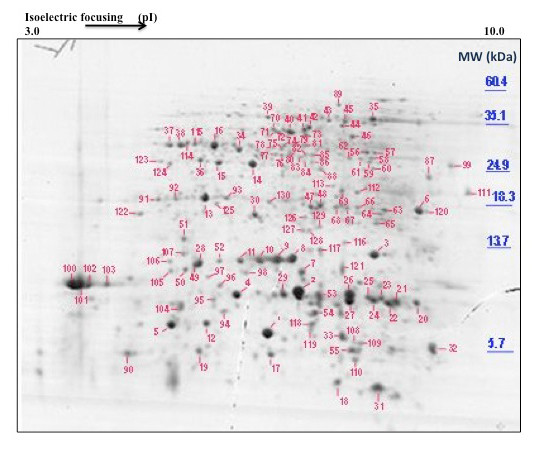
Figure 1.

Typical 2D gel electrophoresis separation of polypeptides in L. albus phloem exudate. Phloem exudate was collected from the vasculature of developing fruits and the inflorescence raceme. 1 mg of protein was separated and stained using colloidal Coomassie Brilliant Blue G250. Protein spots were excised from the gel, digested with trypsin and analysed by partial sequence determination by MS/MS and subsequently identified using database searches. The positions of molecular mass markers are shown to the right of the figure and the pH gradient is indicated at the top of the gel.
