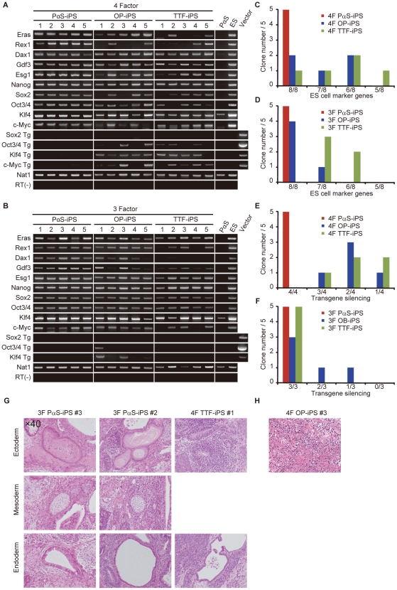
Figure 2. Gene expression and in vivo differentiation of iPS cells.
RT-PCR of 4F-iPS cells (A) and 3F-iPS cells (B) for ES-cell marker genes, including Eras, Rex1, Dax1, Gdf3, Esg1, Nanog, Oct3/4, and Sox2 and to detect silencing of the transgenes. C, Number of ES-cell marker genes expressed using 4F induction. D, Number of ES-cell marker genes expressed using 3F induction. E, Transgenes silenced in 4F induction. F, Transgenes silenced in 3F induction. G, Teratomas from iPS cells, transplanted subcutaneously into nude mice. After 4-6 weeks, the teratomas were analyzed histologically with haematoxylin and eosin staining. TTF-iPS 4F1 did not differentiate mesoderm. H, Undifferentiated cells from 4F OP-iPS #3.

