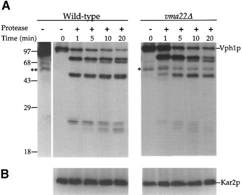
Fig. 3. Partial proteolysis of Vph1p in wild-type and vma22Δ cells. Microsomes were prepared from wild-type (SNY28) and vma22Δ (KHY38) cells and treated for various times with subtilisin (0.8 μg/ml). Precipitated proteins were resolved by SDS–PAGE and immunoblots probed with either anti-Vph1p (A) or anti-Kar2p (B) antibodies. The single asterisk (*) indicates a non-specific band present in vma22Δ immunoblots and ** indicates the same non-specific band present in wild-type immunoblots when overexposed.

An official website of the United States government
Here's how you know
Official websites use .gov
A
.gov website belongs to an official
government organization in the United States.
Secure .gov websites use HTTPS
A lock (
) or https:// means you've safely
connected to the .gov website. Share sensitive
information only on official, secure websites.
