Fig. 6.
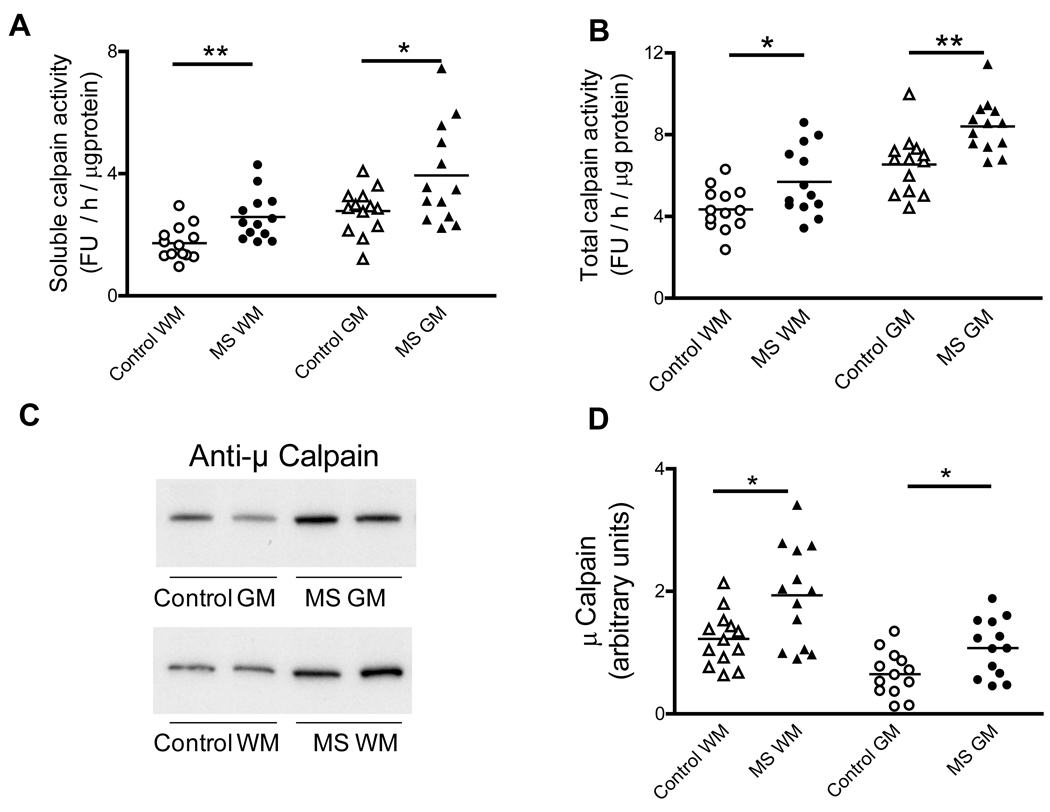
Calpain activity and levels are increased in MS. Aliquots of the cerebral homogenates from control and MS patients were used to determine the soluble (active) (A) and total calpain activity (B) using a fluorescent peptide substrate as described in “Materials and Methods”. Enzyme activity values are expressed as fluorescence units (FU) / hour / µg protein. (C) Representative immunoblots from homogenates of brain WM and GM areas of control and MS patients developed with antibodies against µ-calpain. (D) The relative levels of µ-calpain were calculated by dividing the band intensity on the western blot by that of the corresponding coomassie blue stained lane. Each point represents a patient and the horizontal bar is the average. *p<0.05, **p<0.01. Other symbols are as in Fig. 1.

