
Fig. 2.
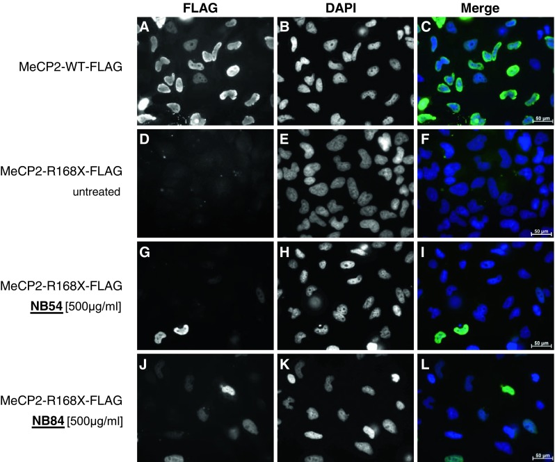
Nuclear localization of readthrough full-length MeCP2-FLAG fusion protein in drug-treated HeLa cells. Immunofluorescence of HeLa cells transfected with MeCP2-WT-FLAG- (a–c) compared to MeCP2-R168X-FLAG-expressing HeLa cells cultured under untreated (d–f) and drug-treated (g–i 500 μg/ml NB54; j–l 500 μg/ml NB84) conditions. Treatment was performed for 24 h. Localization of FLAG fusion proteins (left column) was visualized by using a monoclonal anti-FLAG antibody and immunofluorescence microscopy. Because the fusion protein was C-terminally FLAG-tagged, only full-length MeCP2 proteins were detected. The nuclei were counterstained with DAPI (middle column). Right column MeCP2-WT-FLAG expression as well as treated MeCP2-294-FLAG overlaps with DAPI staining, indicating a nuclear localization. Scale bars are 50 μm