
Fig. 3.
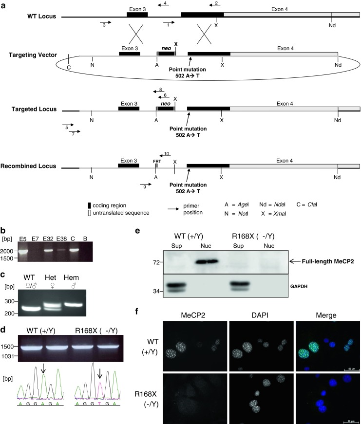
Gene targeting and Flp-mediated recombination at the Mecp2 locus. The diagram shows the wild-type Mecp2 genomic locus, the targeting vector, and the targeted locus before and after Flp-mediated recombination. The 5′ and 3′ regions of homology are shown in light gray. Small arrows indicate primers used to confirm gene targeting and Flp-mediated recombination, and to screen the genotype (a). Homologous recombination was verified by nested PCR of genomic DNA of the selected ES cell clones; primers 5/6 and 7/8 were as shown in a. The 1.9-kb band is from the mutated allele. The primer sets were designed not to amplify the wild-type Mecp2 gene. E5, E7, E32, and E38 are names of ES clones, while c and b indicate a plasmid DNA control demonstrating the mutant PCR fragment and blank. Primer set 9 and 10 were used for the genotyping PCR after FLP recombination (c). The 313-bp band represents the mutant allele (Hem hemizygous male, Het heterozygous female) containing the remaining FRT site, while the 265-bp band represents the wild-type allele (WT wild type). To confirm the presence of Mecp2 mRNA in the brain of two wild-type (WT (+/Y)) and two R168X (−/Y) mutant male mice, total RNA was extracted from brain of male wild-type and mutant mice and used for cDNA synthesis and RT-PCR (d). Western blot analysis (e) and immunofluorescent staining (f) demonstrated the absence of wild-type MeCP2 protein in nuclear extracts of hemizygous brains (Sup supernatant, Nuc nuclear extract) and in nuclei of mouse ear fibroblast using an antibody recognizing the N terminus of MeCP2. Note that no additional truncated protein was detected