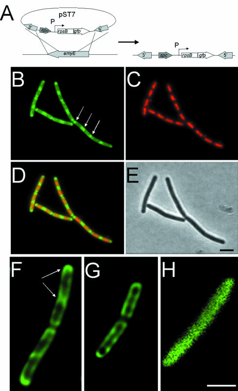
Fig. 4. Ribosome distribution in exponentially growing and stationary phase cells. (A) Construction of the fusion strain 1049 by insertion of pST7 into the chromosome. Integration of pST7 into the amyE gene allows induction of rps–gfp expression from the xylose-inducible promoter (P). (B–D) Distribution of ribosomes in strain 1049 grown in CH medium. (B) RpsB–GFP image false coloured green. Polar ribosomal concentrations are indicated with arrows. (C) DAPI image false coloured red. (D) Image overlays. (E) Phase contrast image. (F–H) Image slices taken from the middle of exponentially growing (F) and stationary phase (G) strain 1049, and exponentially growing strain 1758 (H). The image in (H) has been magnified 2.5 times as it was obtained without the optovar used to acquire the images in (F) and (G), due to the lower signal intensity in this sample. Scale bar, 2 μm.

An official website of the United States government
Here's how you know
Official websites use .gov
A
.gov website belongs to an official
government organization in the United States.
Secure .gov websites use HTTPS
A lock (
) or https:// means you've safely
connected to the .gov website. Share sensitive
information only on official, secure websites.
