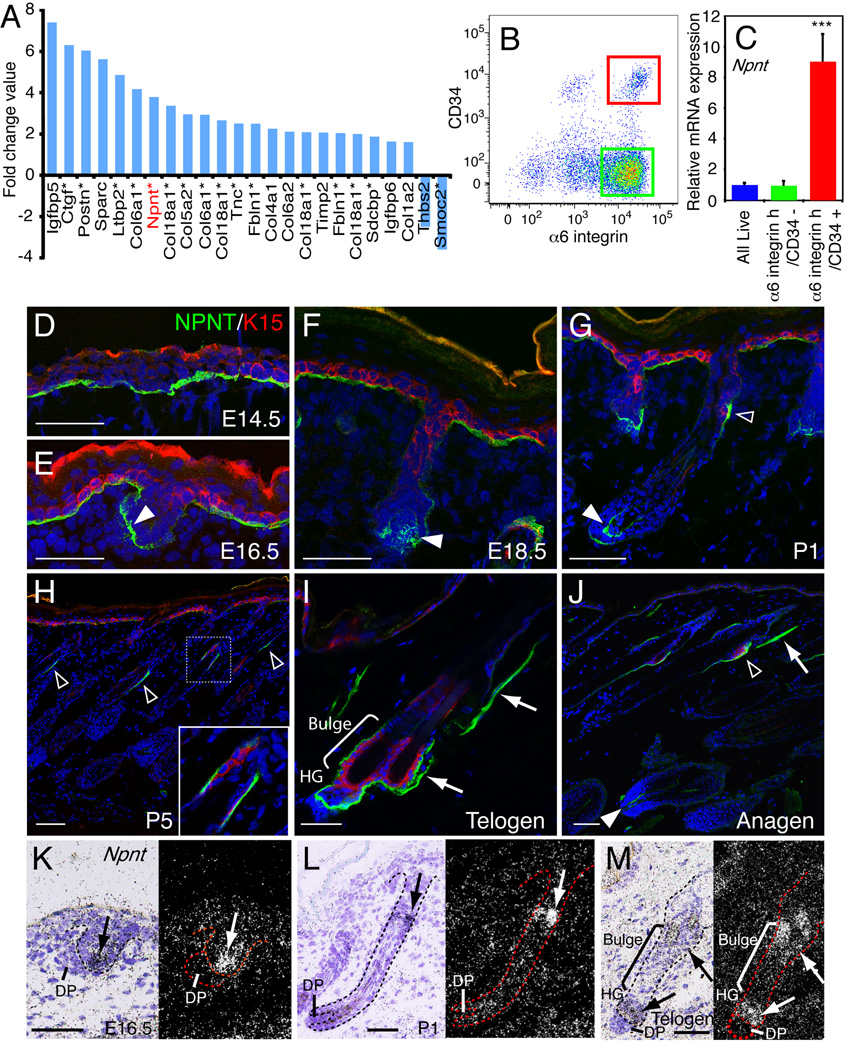
Figure 1. Nephronectin expression in skin.
(A) ECM genes that are upregulated or downregulated in bulge stem cells relative to other basal keratinocytes, ranked based on log2 fold change value (see Table S1). Asterisks indicate the genes that are also upregulated or downregulated in mouse label retaining cells (Tables S2, S3). Some genes are listed more than once due to their multiple spots on the array. (B, C) Adult telogen dorsal keratinocytes were FACS-sorted according to α6 integrin and CD34 expression (B). Bulge stem cells (red gate; α6 integrinhigh/CD34+), non-bulge basal stem cells (green gate; α6 integrinhigh/CD34−) and all live basal cells were sorted and Npnt mRNA levels were determined by Q-PCR (C). Data are mean ± SEM from three mice. (D–J) Sections of E14.5 (D), E16.5 (E), E18.5 (F), P1 (G), P5 (H), adult telogen (I) and anagen (J) skin were immunostained for nephronectin (NPNT; green) and bulge stem cell marker K15 (red), with DAPI counterstain (blue). Note nephronectin deposition in hair germ (white arrowheads), bulge (open arrowheads) and APM (arrows). (K–M) In situ hybridization with Npnt probe on E16.5 (K), P1 (L), and adult telogen skin (M). Arrows indicate the strong nephronectin expression. Scale bars: 50 µm. See also Figure S1.

