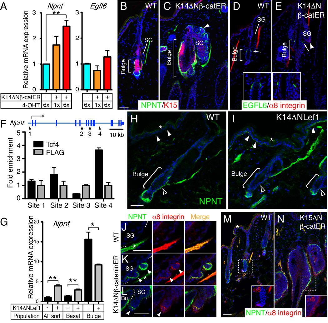
Figure 6. Regulation of regional nephronectin-α8 integrin interaction by Wnt/β-catenin signalling.
(A) Q-PCR of Npnt and Egfl6 mRNA in keratinocytes isolated from skin of wild type (ΔNβ-catER −) and K14ΔNβ-cateninER mice (ΔNβ-catER +) that had been treated with 4-OHT for the number of times indicated. Data are means ± SEM from three mice. (B–E) Dorsal skin of wild type and K14ΔNβ-cateninER mice treated 6× with 4-OHT and immunostained for nephronectin (green; B, C) or EGFL6 (green; D, E) and K15 (red; B, C) or α8 integrin (red; D, E), with DAPI counterstain (blue). Arrowheads indicate ectopic hair follicle arising from sebaceous gland (SG). Arrows indicate EGFL6- staining. Inserts show higher magnification views of EGFL6 staining around the bulge. (F) ChIP using antibodies against Tcf4 and FLAG in K14ΔNβ-cateninER keratinocytes. Primers surrounding four conserved putative binding sites for Lef/Tcfs at the Npnt locus were used to detect the precipitated DNA fragments. Data are means ± SEM of two independent experiments. (G) Q-PCR of mRNA from FACS-isolated bulge, non-bulge (basal) and total basal (All sort) keratinocytes from wild type and K14ΔNLef1 adult telogen skin. Data are means ± SEM from three mice. (H, I) Wild type and K14ΔNLef1 skin immunostained for nephronectin (green) with DAPI counterstain (blue). Basement membrane of interfollicular epidermis (white arrowheads) and bulge (open arrowheads) is indicated. (J–N) Sections of wild type (J, M), K14ΔNβ-cateninER (K, L) and K15ΔNβ-cateninER (N) skin treated 6× (J–L) or 9× (M, N) with 4-OHT and immunostained for nephronectin (green) and α8 integrin (red), with DAPI counterstain (blue). Arrowheads indicate ectopic hair follicles. Asterisks in J and K indicate arrector pili muscles. Inserts in M, N show magnified images of α8 integrin staining around the bulge. Asterisks in H, I and M indicate non-specific staining. Scale bars: 50 µm. See also Figure S5.

