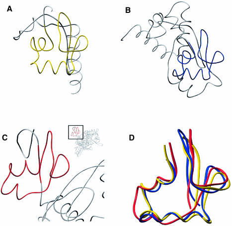
Fig. 5. The αL motif in three different protein structures. The peptide backbones of three structures, ribosomal protein S4, Hsp15 and threonyl–tRNA, are compared. The region highlighted in color is the αL motif that is shared by all three proteins. (A) Hsp15 with its αL motif highlighted in yellow. (B) Ribosomal protein S4 with its αL motif highlighted in blue. (C) Threonyl-tRNA synthetase with its αL motif highlighted in red. (D) Overlay of residues 9–57 of Hsp15 (yellow), 92–141 of ribosomal protein S4 (blue) and 18–59 of threonyl-tRNA synthetase (red).

An official website of the United States government
Here's how you know
Official websites use .gov
A
.gov website belongs to an official
government organization in the United States.
Secure .gov websites use HTTPS
A lock (
) or https:// means you've safely
connected to the .gov website. Share sensitive
information only on official, secure websites.
