
Figure 3.
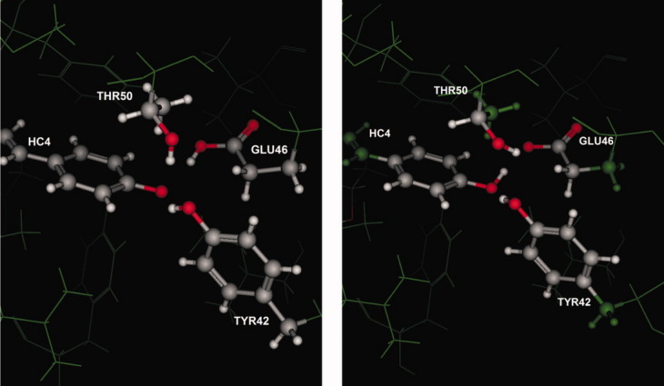
Left is a depiction of the deposited coordinates of 3PYP (photoactive yellow protein) in which Glu46 is neutral and in an unusual conformation and the phenol oxygen of the covalently bound ligand HC4 is apparently anionic. Right is a depiction of the Protonate3D protonation calculation in which the phenol is neutral and the Glu46 is anionic; the hydroxyl of Thr50 is rotated to donate its hydrogen in a polar interaction with the carboxylate.