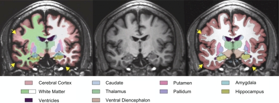
Figure 2.
Coronal MRI image from a male subject with alcoholism showing the results of segmentation from FreeSurfer (left) and CMA (right) in relation to a reference T1 image (center). Arrows (yellow) indicate regions of disagreement between FreeSurfer and CMA regarding the exterior boundary of the brain (lower arrows) and the extent of the white matter into the cortical gyri (upper arrow). There are also diffuse differences in the estimation of sulcal depth.
Abbreviations: CMA, Center for Morphometric Analysis; MRI, magnetic resonance imaging.

