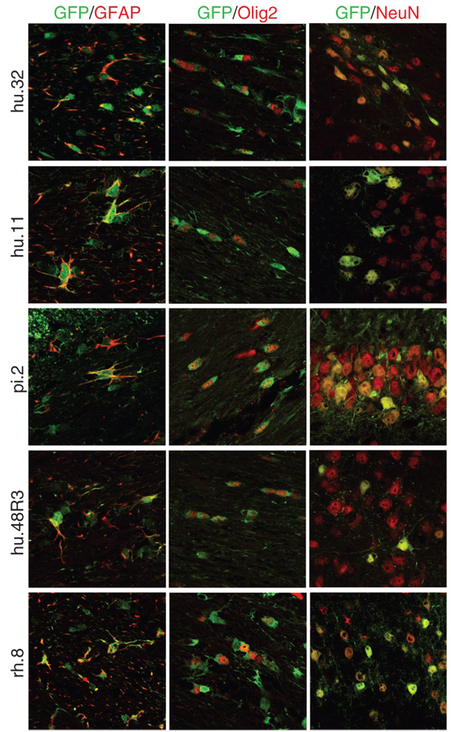
Figure 5. Novel adeno-associated virus (AAV) vectors result in green fluorescent protein (GFP) expression in non-neuronal cells.
GFP-positive sections from brains injected with AAV hu.32, hu.11, pi.2, hu.48R3, and rh.8 were co-labeled with antibodies to the astrocytic marker glial fibrillary acidic protein (GFAP), the oligodendrocytic marker Olig2, or the neuronal marker NeuN. The GFP-positive cells stain green, whereas the cell-specific markers fluoresce red via conjugated secondary antibodies. All pictures were taken using confocal microscopy. GFAP and Olig2 pictures were taken with ×63 magnification and a ×2.3 zoom. NeuN pictures were taken with ×40 magnification and ×2 zoom. Pictures showing co-localization with GFAP or Olig2 were taken of the corpus callosum or external capsule. Pictures showing co-localization with NeuN were taken on the outer edges of the transduced regions and are of the hippocampus (AAV9 or pi.2), the thalamus (hu.32, hu.11, or hu.48R3), or of the cortex (rh.8).

