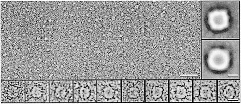
Fig. 2. Formation of large SecYEG structures after incubation with membrane-inserted SecA. SecYEG proteoliposomes were incubated with SecA and AMP–PNP, solubilized and subjected to anion-exchange chromatography. The fraction containing SecYEG was visualized by negative-stain EM, revealing a heterogeneous size distribution (overview, scale bar = 50 nm). A significant fraction of particles had a size of 10–12 nm, a 4–6 nm central stain-filled cavity and a squarish shape (bottom inserts, scale bar = 10 nm). Major class averages (200–400 particles) were generated by single particle analysis (right inserts, scale bar = 5 nm).

An official website of the United States government
Here's how you know
Official websites use .gov
A
.gov website belongs to an official
government organization in the United States.
Secure .gov websites use HTTPS
A lock (
) or https:// means you've safely
connected to the .gov website. Share sensitive
information only on official, secure websites.
