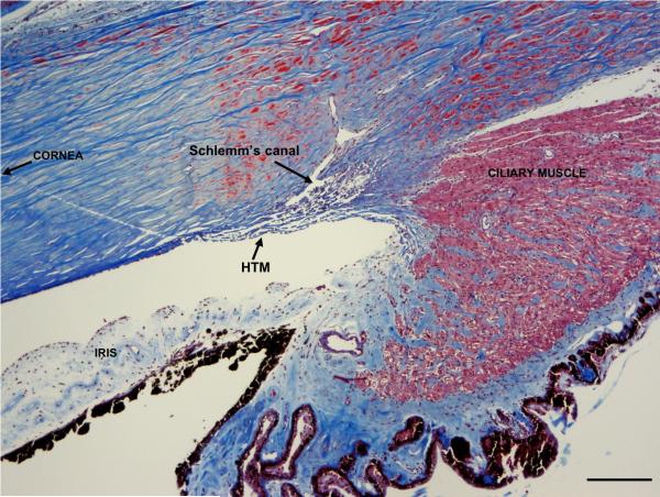
FIGURE 1.
Human Trabecular Meshwork. The HTM is located at the inner corneoscleral junction, near the base of the iris. It is composed of collagen beams (blue) lined by trabecular meshwork cells forming a 3-dimensional “sieve”-like network. This structure is responsible for determining outflow facility, as it directly abuts Schlemm's canal. Ciliary muscle fibers (stained red) insert on the posterior aspect of the meshwork. Masson's trichrome stain, scale bar 200 μm.

