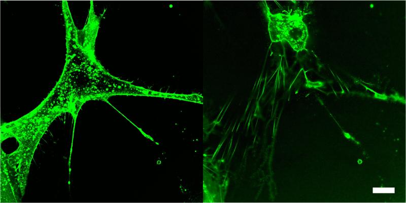
FIGURE 4.
Confocal images of a live HTM cell adhered to glass, before (left) and 30 minutes after exposure to Lat-B (right). Cell membrane was stained with fluorescent wheat germ agglutinin. During collapse of the cell body, delicate retraction fibers coated in WGA, maintain adhesion with underlying substrate. Scale bar 20 μm.

