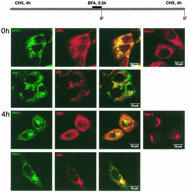
Fig. 10. Effect of BFA on the distribution of retained Kd molecules in the absence of gp40. m152 transfectants were incubated with cycloheximide for 4 h, then BFA was added for 0.5 h. Subsequently, cells were washed three times in the presence of cycloheximide (CHX) before incubation was continued for another 4 h. Double staining was performed at 0 and 4 h after BFA release with mAb against Kd (SF1.1.1., green), polyclonal antibodies against the ER marker calnexin (CNX, red) and the ERGIC marker p58 (red), respectively. BFA-induced changes of the Golgi were followed with polyclonal antibody against ManII (right panels).

An official website of the United States government
Here's how you know
Official websites use .gov
A
.gov website belongs to an official
government organization in the United States.
Secure .gov websites use HTTPS
A lock (
) or https:// means you've safely
connected to the .gov website. Share sensitive
information only on official, secure websites.
