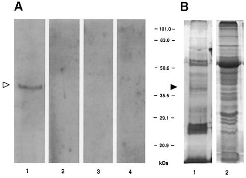
Fig. 1. Detection of TMV MP-interacting protein in cell wall fraction by immunorecognition of bound MP. (A) Renatured blot overlay assay. Lane 1, cell wall fraction blot incubated with MP; lane 2, cell wall fraction blot incubated with buffer alone; lane 3, empty membrane incubated with TMV MP; lane 4, soluble fraction blot incubated with TMV MP. (B) Protein content of the cell wall and soluble fractions of tobacco leaf tissue. Both lanes represent protein extract derived from 10 mg of fresh tobacco leaf tissue. Lane 1, silver-stained cell wall proteins; lane 2, Coomassie blue-stained soluble proteins. The positions of the 38 kDa TMV MP-interacting protein on the blot (open arrowhead) and on stained SDS–polyacrylamide gels (filled arrowhead) are indicated. The numbers between panels indicate molecular mass standards in kDa.

An official website of the United States government
Here's how you know
Official websites use .gov
A
.gov website belongs to an official
government organization in the United States.
Secure .gov websites use HTTPS
A lock (
) or https:// means you've safely
connected to the .gov website. Share sensitive
information only on official, secure websites.
