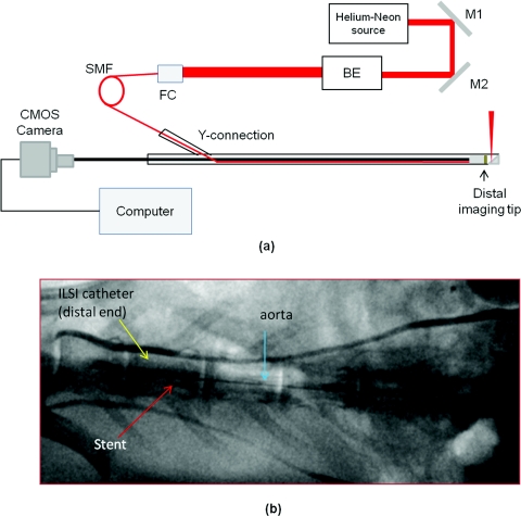
Figure 2.
(a) Schematic of ILSI system. This system incorporates a Helium-Neon light source, an optical setup to couple the laser into the catheter and a high frame-rate camera, BE: beam expander, FC: fiber coupler, M: mirror, SMF: single mode fiber. (b) Fluoroscopic snapshot of the rabbit aorta showing the embedded stent and the ILSI catheter. Two radio-opaque marks are seen on the distal end of the ILSI catheter.

