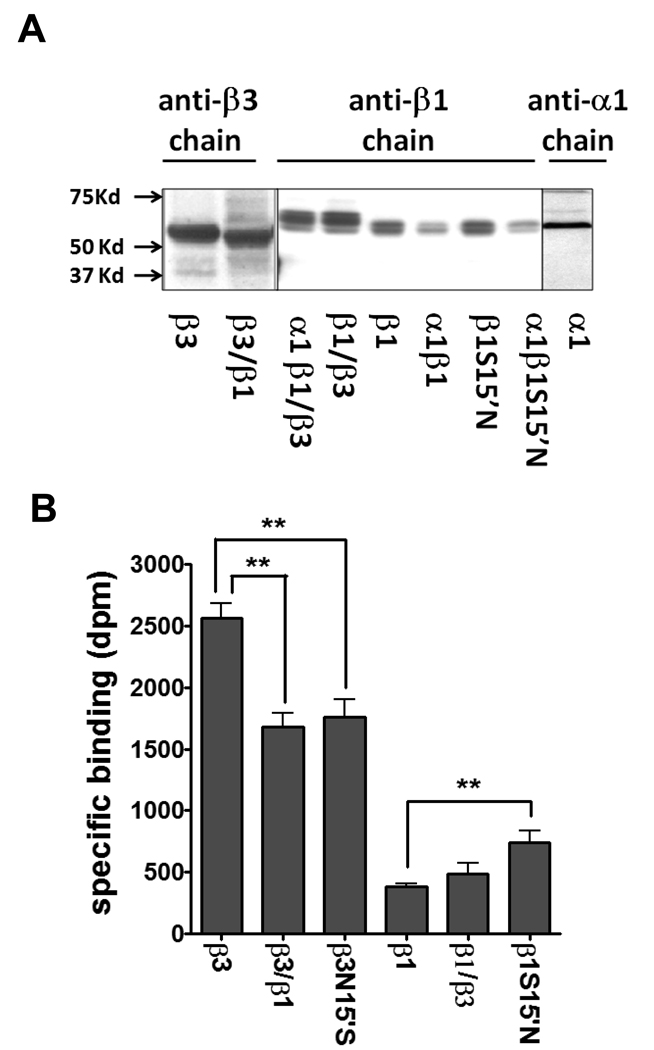
Fig. 3.
Western blotting and [3H]EBOB binding activity of expressed receptors. (A) Individual subunits (α1, β1, β3, β1S15’N, β3/β1 and β1/β3) and co-expressed with α1 subunit (β1, β1/β3 and β1S15’N subunits). Both β3/β1 and β1/β3 are recognized by the anti-β3 chain and anti-β1 chain antibodies, respectively. β3/β1 shows a lower size than β3 WT and β1/β3 a larger size than β1 WT, indicating new proteins are generated. (B) Effect of site-specific mutations and chimeras on specific binding of [3H]EBOB. The data for plotting are given in Table S1 part A. ** P<0.01.

