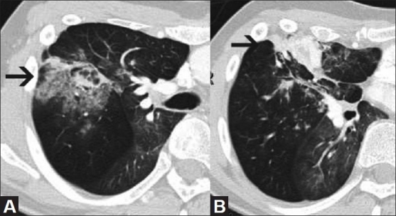
Figure 1(A, B).

CT scan of the chest at two contiguous levels shows areas of bronchiectasis surrounded by air space opacities (arrow in A) representing hemorrhage within the consolidated right middle lobe, extending to the pleural surface (arrow in B)
