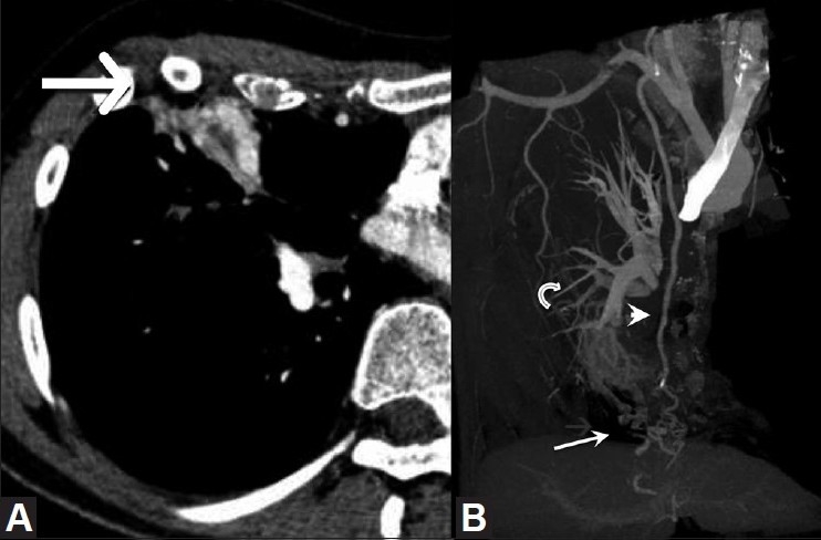
Figure 2.

(A, B): Contrast-enhanced CT scan of the chest (A) demonstrates hypervascularity involving the consolidated right middle lobe (arrow). Bone-subtracted, maximum-intensity projection (B) shows a complex right middle lobe vascular malformation (arrow) supplied by the right internal mammary artery (arrowhead) with drainage into the right pulmonary artery (curved arrow)
